Fig. 7.
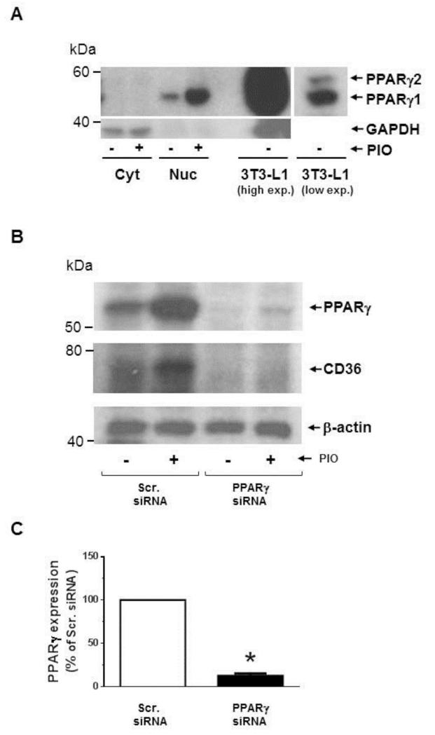
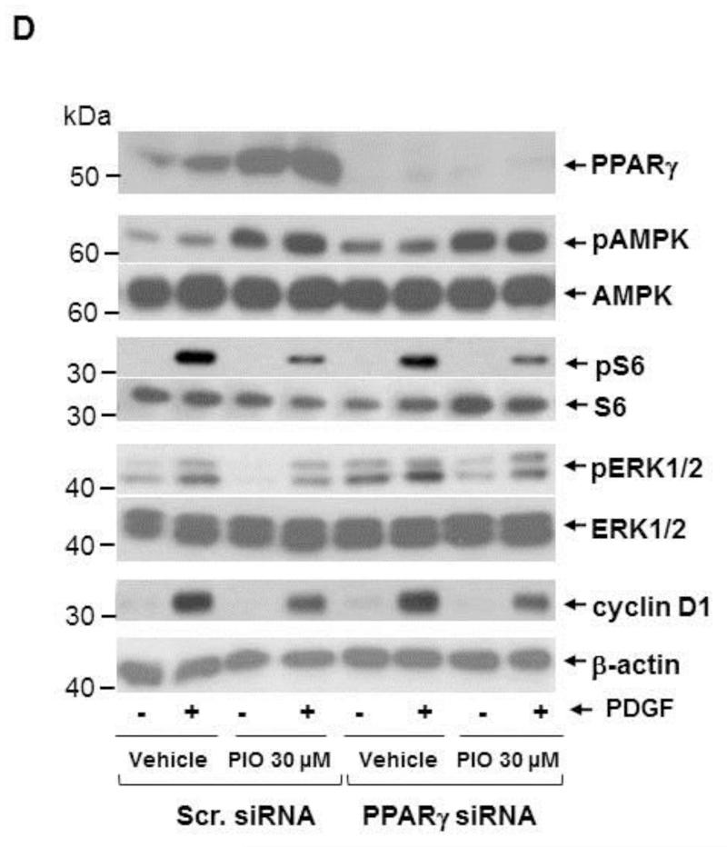
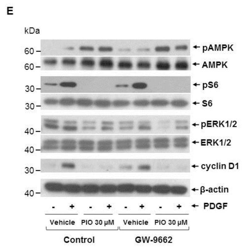
Effects of PPARγ downregulation or PPARγ inhibition on PIO-induced changes in the phosphorylation of AMPK, S6, and ERK1/2, and cyclin D1 expression in VSMCs. (A) Serum-deprived VSMCs were treated with PIO (30 μM, 48 hr) or vehicle control. Subsequently, nuclear (Nuc.) and cytoplasmic (Cyt.) proteins (20 μg) were extracted for immunoblot analysis of PPARγ. Differentiated 3T3-L1 cell lysate (2 μg) was used as a positive control for the expression of PPARγ. Immunoblots shown are representative of n = 3. (B-D) VSMCs were transfected with scrambled (Scr.) or PPARγ siRNA followed by maintenance in culture for 48 hr. Subsequently, VSMCs were pretreated with PIO (30 μM, 30 min) or vehicle control followed by exposure to PDGF (30 ng/ml, 48 hr) under serum-deprived conditions. The respective VSMC lysates were then subjected to immunoblot analysis using primary antibodies specific for PPARγ, CD36, pAMPK, pS6, pERK1/2 and cyclin D1. *p < 0.05 compared with scr. siRNA. Immunoblots shown are representative of n = 3. (E) Serum-deprived VSMCs were pretreated with PPARγ inhibitor (GW-9662; 10 μM, 30 min) followed by exposure to PIO (30 μM, 30 min) or vehicle control and subsequent exposure to PDGF (30 ng/ml, 48 hr). VSMC lysates were then subjected to immunoblot analysis for pAMPK, pS6, pERK1/2 and cyclin D1. Immunoblots shown are representative of n = 3.


