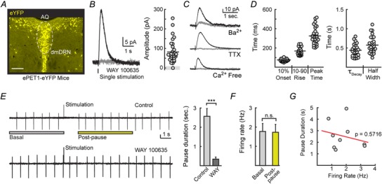
Figure 1. Electrical stimulation evokes 5‐HT1A‐IPSCs that drive pauses in DRN serotonin neuron firing .

A, eYFP, driven by the PET1 enhancer, selectively labels serotonin neurons. Shown is eYFP immunoreactivity in a coronal brain slice obtained from ePET1‐eYFP mice containing the dorsal raphe nucleus (DRN). AQ, cerebral aqueduct; dmDRN, dorsomedial dorsal raphe nucleus. Scale bar represents 200 μm. B, left, averaged 5‐HT1A‐IPSCs from a dorsal raphe neuron evoked by a single stimulation in control conditions (black) and in the presence of the 5‐HT1A antagonist WAY 100635 (200 nm; grey). Whole‐cell recordings were voltage‐clamped to −60 mV. Right, IPSCs could be evoked in 30/31 DRN serotoninergic neurons examined. C, average traces of IPSCs recorded in 200 nm TTX, calcium‐free ASCF, and 200 μm barium. D, quantification of the rise and decay kinetics of evoked IPSCs. E, cell‐attached recordings made in 3 μm phenylephrine to mimic in vivo excitatory drive. Representative traces (left) and quantification (right) demonstrating that evoked serotonin release drives a pause in firing that is absent in the 5‐HT1A‐receptor antagonist WAY 100635 (200 nm). Vertical scale bar represents 25 pA. F, firing rates measured over 5 s before stimulation (basal) and over 5 s after firing had resumed (post‐pause) were identical. G, Pearson correlation demonstrating that the duration of the evoked pause was independent of the baseline firing rates (n = 9).
