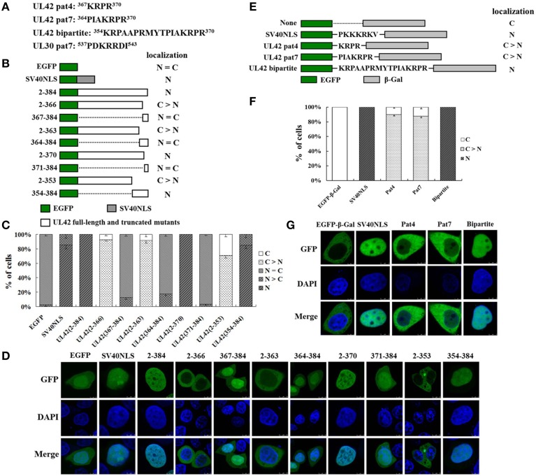
Figure 2.
UL42 contains a functional and transferable bipartite NLS that mediates its nuclear localization. (A) The predicted NLSs in the PRV UL42 and UL30 sequences identified with the PSORT II software program. The single-letter amino acid code is used. The superscript numbers indicate the corresponding amino acid positions within the protein sequence. (B) Schematic representation of EGFP, EGFP–SV40NLS, and full-length EGFP–UL42 and their truncated mutant derivatives, which were used to identify the putative NLS in UL42. The localization of various EGFP-expressing fusion proteins was categorized into five different patterns: N, exclusively nuclear; N > C, more nuclear than cytoplasmic; N = C, diffuse; C > N, more cytoplasmic than nuclear; and C, strictly cytoplasmic. The localization results are summarized on the right. (C) For each construct, 100 EGFP-expressing cells were scored from independent transfections in three repeated experiments and the relative percentages of the different subcellular localization categories of the fusion constructs were calculated. N, exclusively nuclear; N > C, more nuclear than cytoplasmic; N = C, diffuse; C > N, more cytoplasmic than nuclear; C, strictly cytoplasmic. (D) Representative localization images of various UL42–EGFP fusion protein constructs. Localization of the fusion proteins was analyzed as the fluorescent EGFP signal with confocal microscopy, and the DNA was stained with Hoechst reagent. The merged GFP and DAPI signals are shown. The image for each construct is representative of three independent transfection experiments. (E) Schematic representation of the classical SV40 TAg-NLS and the predicted UL42 pat4, pat7, or bipartite NLS fused between EGFP and β-Gal. “None” indicates that no specific motif was fused between these two reporter proteins. The localization of these constructs is summarized on the right. N, exclusively nuclear; C > N, more cytoplasmic than nuclear; C, strictly cytoplasmic. (F) For each construct, 100 EGFP-expressing cells were scored after independent transfections in three repeated experiments and the relative percentages of the different subcellular localizations of the fusion constructs were estimated. N, exclusively nuclear; C > N, more cytoplasmic than nuclear; C, strictly cytoplasmic. (G) Representative localization images of various EGFP–β-Gal fusion proteins containing the specific NLS. Localization of the fusion proteins was analyzed as the fluorescent EGFP signal using confocal microscopy, and the DNA was stained with Hoechst reagent. The merged GFP and DAPI signals are shown. The image for each construct is representative of three independent transfection experiments.

