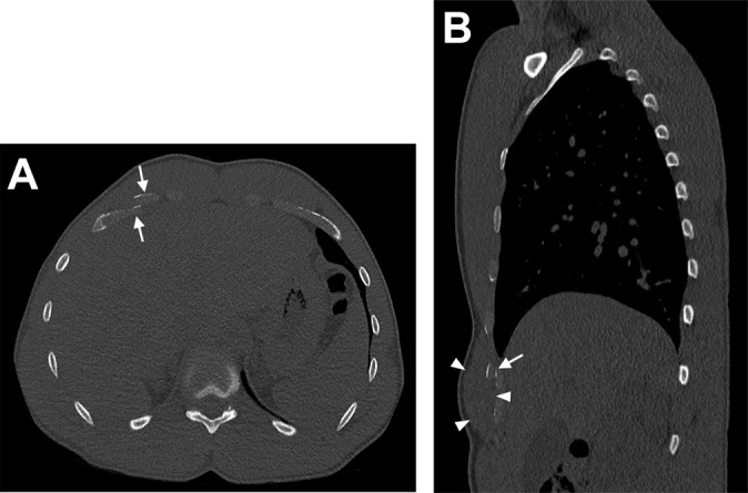
Figure 2.

Case 2. (A) Transverse, noncontrast computed tomography (CT) image at the level of the right sixth rib showing fracture and overlap of the sixth costal cartilage (arrow). Other nondisplaced fractures not shown. (B) Sagittal reformation CT showing costal cartilage fracture (arrow) and adjacent soft tissue swelling involving adjacent musculature (arrowheads).
