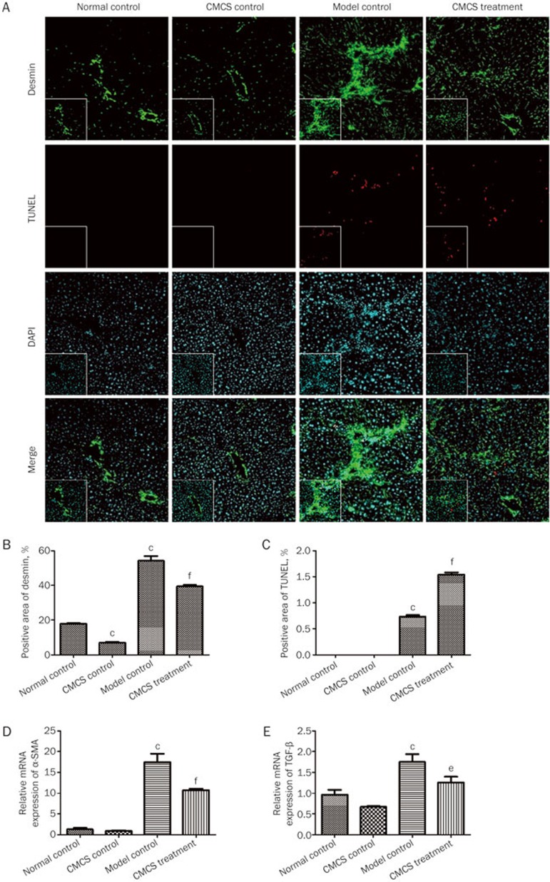
Figure 3.
CMCS treatment restrains HSCs activation in mice with CCl4-induced liver fibrosis. (A) Liver sections were subjected either to immunohistochemical staining for desmin (green) for detection of HSCs, the TUNEL assay to detect cell apoptosis (red) or blue-fluorescent DAPI nucleic acid staining (magnification, 200×). Desmin-positive (B) and TUNEL-positive (C) areas were quantified. (D and E) Relative mRNA expression levels of α-SMA (D) and TGF-β (E) were assayed by RT-qPCR. Values represent mean±SD (n=10). bP<0.05, cP<0.01 versus normal control group; eP<0.05, fP<0.01 versus model control group.

