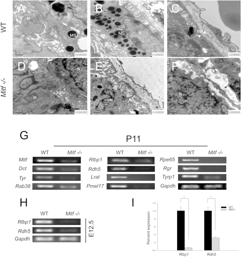
Figure 1. Abnormal RPE ultrastructures and decreased expression of visual cycle genes in Mitf−/− mice.
(A–F) P0 (A–E) and P20 (C,F) RPE ultrastructures of WT and Mitf−/− mice revealed by transmission electron microscopy (TEM). (A,D) show the interface between RPE and photoreceptor cells in WT and Mitf−/− mice, respectively. The RPE in Mitf−/− displayed thickened and shortened microvilli as compared to WT. The microvilli in the apical surface of the RPE are indicated by MV. B and E show the basal part of the RPE in WT and Mitf−/−, respectively. Bruch membrane was discontinuous and disintegrated in Mitf−/−. (C,F) show the basal infoldings of RPE in WT and Mitf−/− mice, respectively. The basal infoldings of RPE were sparse and disordered in Mitf−/− when compared with that of WT. BI, basal infoldings; BM, Bruch’s membrane; Ch, choroid; M, mitochondria; MS; melanosome; MV, microvilli; N, nucleus. (G) RT-PCR analysis of the expression of visual cycle and pigmentation genes in P11 retinas/RPE of WT and Mitf−/− mice. (H) RT-PCR analysis of the expression of Rlbp1 and Rdh5 in E12.5 optic cups of WT and Mitf−/− mice. (I) Real-time RT-PCR analysis of Rlbp1 and Rdh5 expression in WT and Mitf−/− optic cups at E12.5. RNA levels of Rlbp1 and Rdh5 were normalized to those of Gapdh. The experiments were performed in triplicates and the error bars represent the S.E. *indicates P < 0.05. Gapdh was used as internal control.

