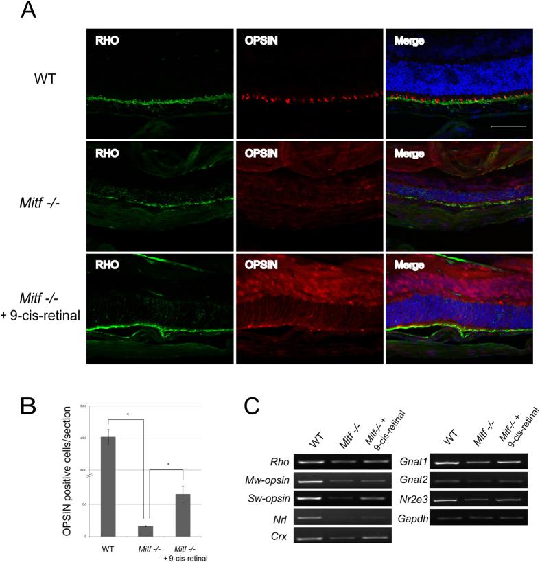
Figure 5. Photoreceptor cells and their related gene expression were maintained in Mitf−/− mice treated with 9-cis-retinal.
(A) RHODOPSIN and OPSIN expression in superior retina of WT and Mitf−/− treated with solvent or 9-cis-retinal. Rod RHODOPSIN was localized to the rod outer segment (ROS), and cone OPSIN was localized to the ONL and the rod outer segment in WT neural retina. In Mitf−/− mice treated with solvent, the RHODOPSIN and OPSIN localization in ROS were decreased and strikingly mislocalized to the cone cell bodies in the ONL. By contrast, RHODOPSIN and OPSIN localization in ROS, especially Rhodopsin, were maintained in Mitf−/− mice treated with 9-cis-retinal. (B) Quantification of OPSIN-labeled cells in retinal ROS of WT, Mitf−/− treated with solvent or treated with 9-cis-retinal. In contrast to WT, the OPSIN-labeled cells in retinal ROS were almost completely lost in Mitf−/− mice treated with solvent, whereas OPSIN-positive cells were partially preserved in Mitf−/− mice treated with 9-cis-retinal. (C) The expression of photoreceptor marker genes in WT, Mitf−/− treated with solvent, and Mitf−/− mice treated with 9-cis-retinal at P22 by RT-PCR. For photoreceptor marker genes, 9-cis retinal administration partially restored S-opsin, M-opsin and Gnat2 expression. Moreover, 9-cis retinal administration also restored expression of the rod-specific genes Rho and Gnat1, as well as genes expressed by both rod and cone cells, including Crx, Nrl, and Nr2e3 expression. Scale bar: 50 μm.

