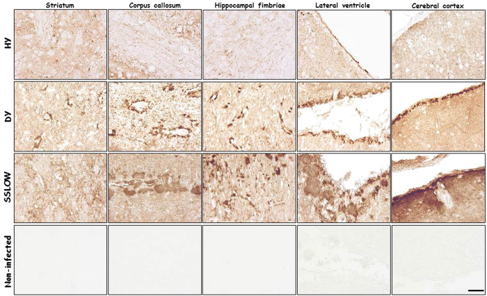
Figure 3. Strain specific PrPres deposition in different brain areas.
Representative pictures of PrP deposition in different brain regions of terminally ill animals infected with HY (n = 5), DY (n = 5), and SSLOW (n = 3) strains. Samples were processed with the 6H4 antibody after PK treatment. Brain sections from a non-infected animal (n = 3) are shown at the bottom panels as a control for staining-specificity against the PK-resistant form of the prion protein. The scale bar (50 μm) at the right-bottom panel is representative of all panels depicted in this figure.

