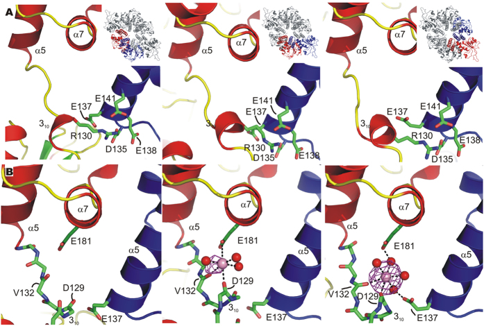
Figure 5. The control elements governing OBD movement in the RepB X-ray structures.
(A) The location of R130 at the OD/OBD interface and of the D and E residues of the N-terminal end of helix α5 that interact with R130 is shown. The insets show the position of the protomers in the C2 hexamer. (B) Views on the region where OBD conformation-dependent metal binding occurs at the different OD/OBDs interfaces of the structure cocrystallised with BaCl2. The protein chain is represented by a cartoon drawing in all panels, except for the interdomain loop region (residues 128–134) containing the 310 helix, for which only a stick representation of the protein backbone is shown. The non-interacting side chains are left out for clarity. The neighbouring protomer is coloured blue and the Ba2+ ions are represented by a pink sphere. The contours represent the Ba2+ anomalous map (see text) contoured at 5σ. Direct and indirect contacts between protein residues and the metal ion, when present, are indicated by dashed lines. The OBD and OD are from two adjacent protomers and are colour-coded as in panels (A).

