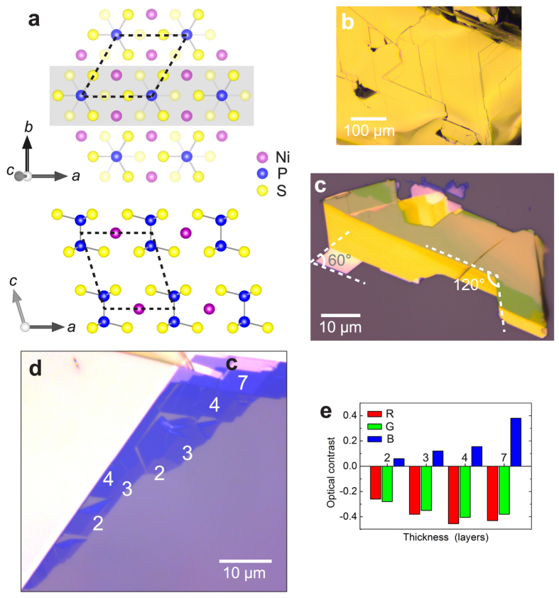
Figure 1. Atomic structure and optical characterization of exfoliated NiPS3.
(a) Schematic crystal structure. View perpendicular to layers, only top layer shown (upper schematic). The atoms contained in the section shaded grey are shown in view parallel to layers (lower schematic). Unit cell (dashed outlines), covalent bonds within (P2S6)4− anions (grey lines). (b) Brightfield microscope image of cleaved bulk NiPS3 samples. (c) Image of NiPS3 flake exfoliated onto oxidized silicon substrate, comprising mainly thick sheets (>10 layers). 60° and 120° angles indicated. (d) Image of thin exfoliated NiPS3 sheets, 2–7 layers indicated. (e) Optical intensity contrast of ultrathin NiPS3 sheets (2–7 layers), evaluated for red, green, and blue color channels separately, with reference to the substrate. (Photos in panels (c) and (d) were acquired at different illumination conditions to maximize visibility of relevant features).

