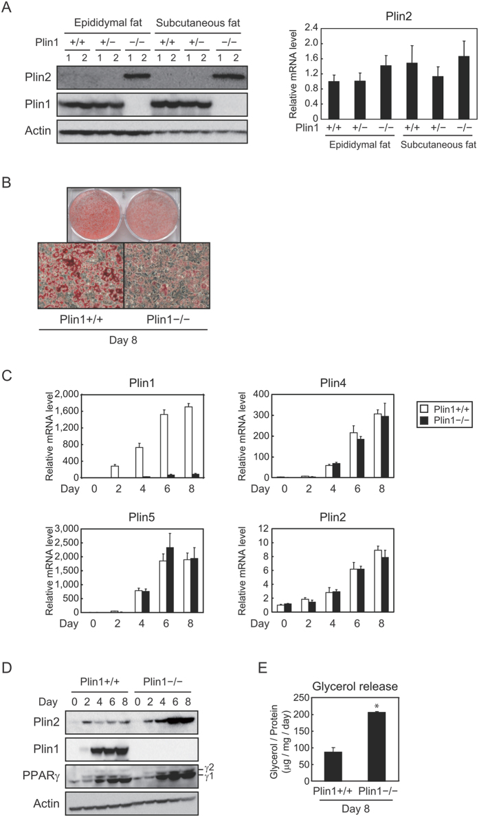
Figure 3. Protein levels of Plin2 are increased during adipocyte differentiation of Plin1 deficient MEFs.
(A) Epididymal and subcutaneous fat pads were isolated from 10–14 weeks-old Plin1+/+, Plin1+/− or Plin1−/− mice fed ad libitum (n = 6, 3 samples were pooled each). Each protein level was detected using Western blot analysis (left) and the Plin2 mRNA level was determined using quantitative RT-PCR and normalized to 18s rRNA levels (right). These data represent the means ± S.D. The assay was performed in triplicate. (B) Differentiated (day 8) MEFs obtained from Plin1+/+ or Plin1−/− mice were stained with Oil Red O. (C) Plin1+/+ and Plin1−/− MEFs were differentiated into adipocytes and harvested every 2 days. Each mRNA level was determined using quantitative RT-PCR and normalized to 18s rRNA levels. These data represent the means ± S.D. The assay was performed in triplicate. (D) Plin1+/+ or Plin1−/− MEFs were differentiated into adipocytes and harvested every 2 days. Each protein level was detected using Western blot analysis. (E) After 8 days of differentiation, glycerol levels contained in the supernatant were determined and normalized to total cellular protein levels as in the Materials and Methods. These data represent the means ± S.D. The assay was performed in triplicate. *P < 0.01.

