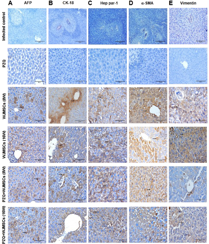
Figure 3.
Photomicrographs of immunohistochemical staining (immunostain, DAB, X400) of (A) human alpha fetoprotein (AFP), (B) cytokeratin-18 (CK-18), (C) Hep par-1, (D) alpha smooth muscle actin (α-SMA), and (E) vimentin in liver sections of control and treated groups. No expression was observed in infected control and praziquantel (PZQ)-treated groups, whereas newly-formed liver-like cells in groups treated with Wharton’s jelly-derived mesenchymal stem cells (WJMSCs) showed positive expression (brown cytoplasmic discolouration) for AFP, CK-18, and Hep par-1. Positive expression of α-SMA and vimentin was observed only in scattered liver-like cells in groups treated with WJMSCs with the least expression observed in the groups which received WJMSCs combined with PZQ.

