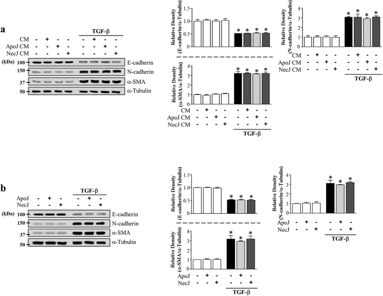
Figure 3. Effect of direct exposure of LA-4 cells to apoptotic cells on TGF-β1-induced EMT.
(a) LA-4 cells were stimulated with apoptotic (ApoJ) or necrotic (NecJ) Jurkat cells for 20 h. Conditioned medium (CM) was added to LA-4 cells in the presence or absence of 10 ng/ml TGF-β1. (b) LA-4 cells were directly exposed to ApoJ or NecJ cells in the presence or absence of 10 ng/ml TGF-β1. (a,b) After 72 h, western blot analysis of EMT markers in LA-4 cells. Values represent the mean ± s.e.m. of three independent experiments. *P < 0.05 compared with control; +P < 0.05 as indicated.

