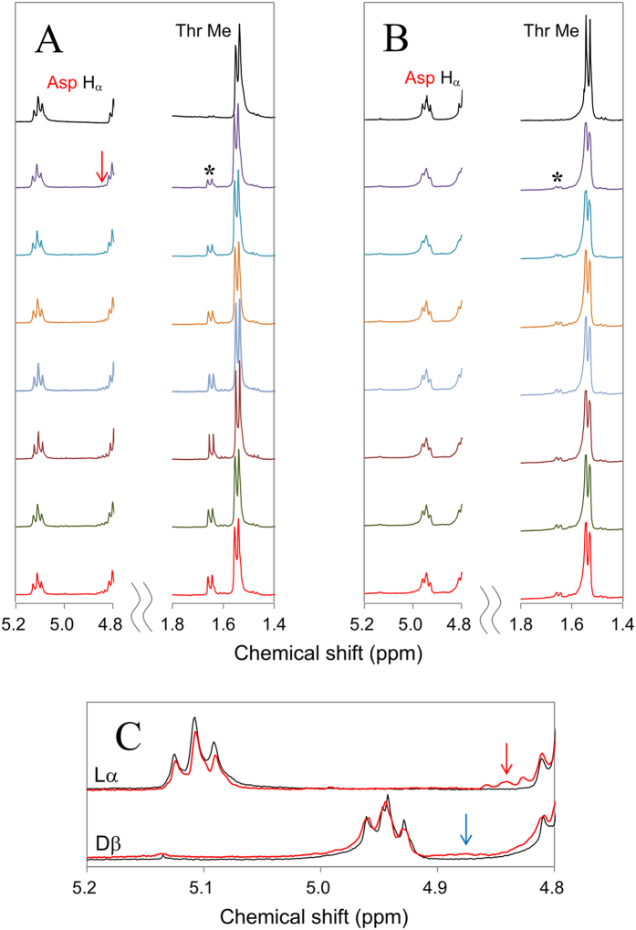
Figure 5. Real time 1H-NMR spectra of αB-crystallin 61–67 analog containing L-α- and D-β-Asp62.
Panel (A) illustrates 1H-NMR spectra for the peptide containing L-α-Asp62 immediately after and 16, 20, 24, 28, 32, 35, and 38 h after heating at 70 °C (from top to bottom). Panel (B) shows 1H-NMR spectra for 61–67 peptide containing D-β-Asp62 immediately after and 23, 26, 30, 32, 34, 36, and 38 h after heating at 70 °C. Each panel shows Asp62 Hα (5.2–4.8 ppm) and Thr63 methyl regions (1.8–1.4 ppm). In panel (C), the Hα region of L-α- and D-β-Asp62 immediately after (in black) and 38 h after heating (in red) is expanded in upper and lower traces, respectively.

