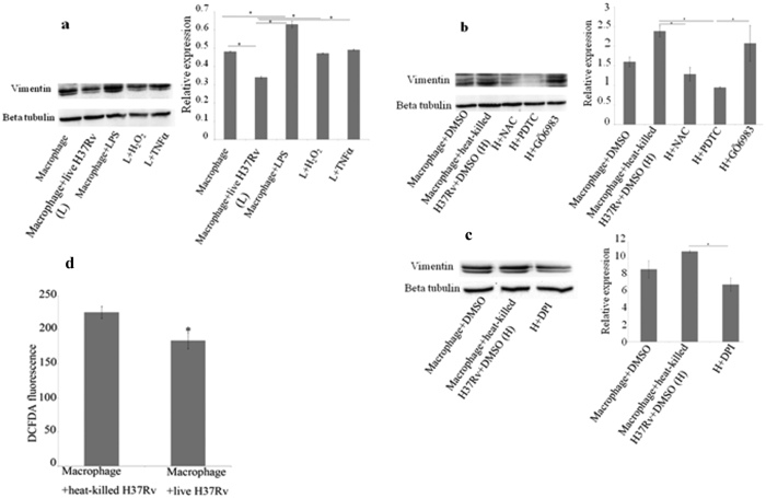
Figure 3. Downregulation of vimentin by Mycobacterium tuberculosis relates to the pathogen’s anti-inflammatory effect on macrophages.
(a) Macrophage monolayers were infected with live H37Rv. Infected macrophages were treated with H2O2 at100 μM and TNFα at10 ng/ml, after 4 h of phagocytosis. Uniinfected macrophages were treated with LPS at1 μg/ml. After 12 h post infection, all the samples were processed for Western blotting and probed with anti-vimentin antibody; β-tubulin was used as loading control. Live mycobacteria reduce vimentin expression and this is restored by H2O2 and TNFα. LPS increases vimentin and is used as a positive control. Values are expressed as mean ± SE, n = 3, *Mean difference is significant at 0.05 level. (b,c) Macrophage monolayers were infected with heat-killed H37Rv. Infected macrophages were treated with NAC at10 mM, PDTC at 50 μM, GÖ6983 at 500 nM and DPI at10 μM, after 4 h of phagocytosis. After 12 h post infection, all the samples were processed for Western blotting and probed with anti-vimentin antibody; β-tubulin was used as loading control. Values are expressed as mean ± SE, n = 3, *Mean difference is significant at 0.05 level. (d) The level of ROS is found to be decreased in live H37Rv-infected macrophages compared to heat-killed H37Rv-infected macrophages after 24 h of infection. Values are expressed as mean ± SE, n = 4 and p = 0.03.

