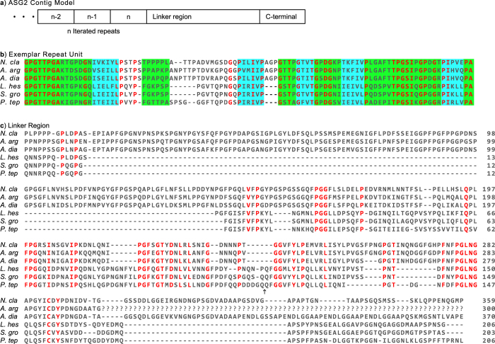
Figure 5. ASG2 from six species.
(a) Schematic of ASG2 architecture inferred from our assembled sequences. Repetitive region contains an undetermined (n) number of repeat units followed by linker and carboxyl (C)-terminal regions. Ellipsis signifies unknown sequence. (b) Alignment of exemplar ASG2 repeat units indicating sequences predicted to form β-sheet (blue shading) or β-turn/random coil structure (green shading). (c) Alignment of linker regions. Amino acids depicted by single letter IUPAC abbreviations. Red indicates amino acids conserved across species and black arrow indicates location where additional sequence from P. tep whole genome assembly contig 63868 was added to our P. tep capture contig. The two sequences were 97% identical over 1874 bp. Abbreviations of species names and GenBank accession numbers are in Table 2.

