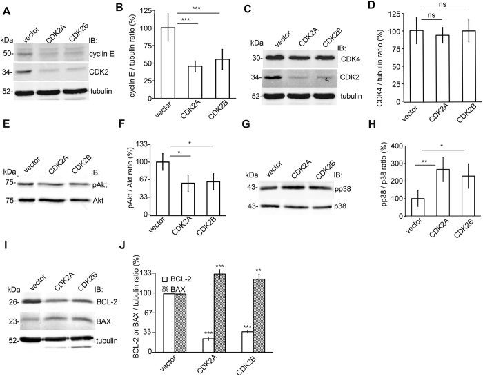
Figure 7. CDK2 knockdown inhibits cyclin E and the antiapoptotic pathways and stimulates the pro-apoptotic pathways in cultured human podocytes.
(A) Immunoblot of cyclin E and CDK2 after knockdown of CDK2 with two different shRNA constructs (CDK2A and CDK2B) in cultured podocytes. Empty vector (vector) served as a control. Tubulin is included as a loading control. (B) Quantification of cyclin E shows that the expression level of cyclin E is downregulated after knockdown of CDK2. (C) Immunoblot of CDK4 after CDK2 knockdown in cultured podocytes. Tubulin is included as a loading control. (D) Quantification of CDK4 shows that the expression level of CDK4 does not change after CDK2 knockdown confirming the specificity of the knockdown. (E) Immunoblot assay of phosphorylated Akt (pAkt) in podocytes after CDK2 knockdown. Total Akt is included as a loading control. (F) Quantification of phosphorylated Akt shows that CDK2 knockdown decreases Akt activity in cultured podocytes. (G) Immunoblot of phosphorylated p38 (pp38) in podocytes after CDK2 knockdown. Total p38 is included as a loading control. (H) Quantification of phosphorylated p38 shows that CDK2 knockdown increases p38 activity in cultured podocytes. (I) Immunoblot of BCL-2 and BAX after knockdown of CDK2 in cultured podocytes. Tubulin is included as a loading control. (J) Quantification of BCL-2 and BAX shows that the expression level of BCL-2 is downregulated and BAX upregulated after knockdown of CDK2. Each experiment was performed three times with three replicates in each experiment. Data are presented as mean ± SD. ns: non significant, *p < 0.05, **p < 0.01, ***p < 0.001 vs. vector group.

