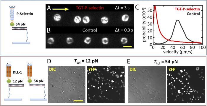
Figure 5. ProG-TGT for P-selectin and Notch receptor.
(A) P-selectin-Fc:ProG-TGTs were assembled and immobilized on the inner surface of a flow chamber where leukocytes were flowed through. A single leukocyte location was captured by consecutive dark field imaging with 3 sec time interval. Yellow arrow indicates flow direction. (B) Single leukocyte location with 0.3 sec time interval on control surface without P-selectin coating. Scale bar: 20 μm. (C) Velocity distribution. HL-60 cells attached onto P-selectin-Fc:ProG-TGTs and show rolling behavior. This resulted in a cell moving rate significantly lower than the rate on control surface. (D) DLL1-Fc:ProG-TGTs were assembled and immobilized on glass surface where Notch activation was tested. Notch receptors were activated on both Ttol = 12 pN surface and Ttol = 54 pN surface after incubation for two days. Notch activation was reported by H2B-YFP expression in CHO-K1 cell nucleus. Scale bar: 50 μm.

