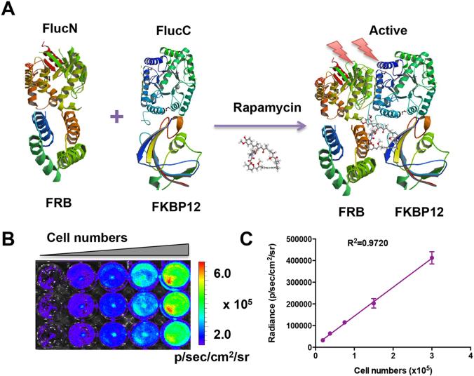
Figure 1.
Characterization of the split luciferase reporter. (A) A crystallographic model of FlucN-FRB and FKBP12-FlucC domains in the reporter system. In the absence of rapamycin, there is no luciferase activity. Upon treatment with rapamycin, the dimerization of FRB and FKBP12 will lead to the activation of the luciferase reporter. The Fluc, FRB and FKBP12 images were adapted from RCSB PDB (www.pdb.org) using ID# 1LUC, 1AUE and 2PPN. (B) Bioluminescence imaging analyzed the linearity of recovered luciferase activity with cell numbers after addition of free rapamycin (20 nM) for 6 h. (C) Linear regression analysis between luminescence intensity and cell numbers.

