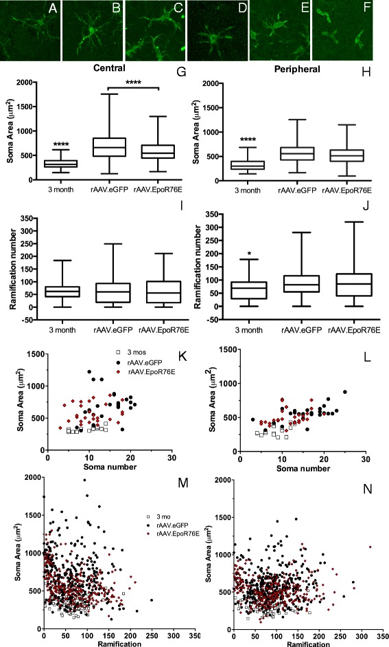
Fig. 3.

Treatment with rAAV.EpoR76E altered microglial morphology. a–f Representative high-magnification confocal micrographs of IBA-1 immunolabeled (green) microglia in 3-month-old (a) and 8-month-old (b–f) retinas. These morphologies were present in retinas from both treatment groups. g, h Box and whisker plots of microglial soma area of cells from the central (g) or peripheral (h) retina, ****p < 0.0001. i, j Box and whisker plots of microglial ramification number for cells from the central (i) or peripheral (j) retina, *p < 0.05. k, l Scatter graphs of microglial soma area versus soma number in the central (k) or peripheral (l) retina. m, n Scatter graphs of microglial soma area versus ramification number in the central (m) or peripheral (n) retina
