Figure 1.

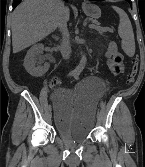
Vertical section of the computed tomography abdomen and pelvis showing the distended bladder (solid arrow head), with diverticula (open arrow), diverticular mouth (double arrow) and right kidney hydronephrosis

Vertical section of the computed tomography abdomen and pelvis showing the distended bladder (solid arrow head), with diverticula (open arrow), diverticular mouth (double arrow) and right kidney hydronephrosis