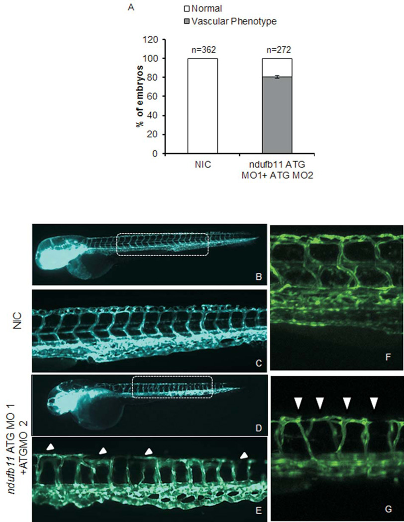
Figure 3.
Microinjection of ndufb11morpholino (MO) in zebrafish embryos leads to vasculature defects. (A) Bar graph showing embryos with normal and defective inter-segmental vessel in non-injected control (NIC) and 400 µM ndufb11ATG MO 1+ ATG MO 2 morpholino injected embryos at 72 hpf. (B–G) Representative images of 72 hpfTg(fli1:EGFP, gata1a: dsRed) zebrafish embryos. (B,C,F) Non-injected control embryos (NIC) with normal vasculature and (D,G,H) ndufb11 MO injected embryos displaying vasculature defect. (B–E) 2.5× magnification. (F,G) 20× magnification. Images are arranged in a lateral view and inset displaying intersegmental vessels from the trunk region. Arrowheads indicate regions with vascular defects.

