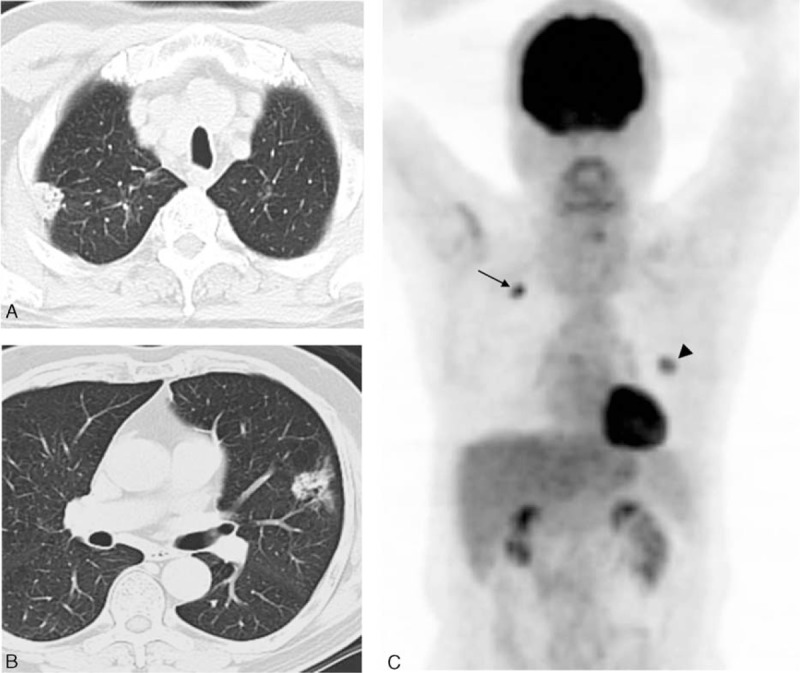
FIGURE 3.

Representative CT and 18F-FDG PET images of a 62-y-old man with 2 synchronous primary lung tumors. (A, B) Axial CT scans in lung window setting showing a 2.0-cm solid nodule in the right upper lobe (RUL) of lung and another 3.1-cm lesion mixed with solid and ground glass components in the left upper lobe (LUL). (C) Whole-body maximum-intensity projection of FDG PET demonstrating a moderately differentiated adenocarcinoma in the RUL with an SUVmax of 9.4 (arrows) and a second primary with similar histology but a different colonel origin in the LUL with an SUVmax of 4.3 (arrowheads). CT = computed tomography, 18F-FDG = 18F-fluorodeoxyglucose, PET = positron emission tomography.
