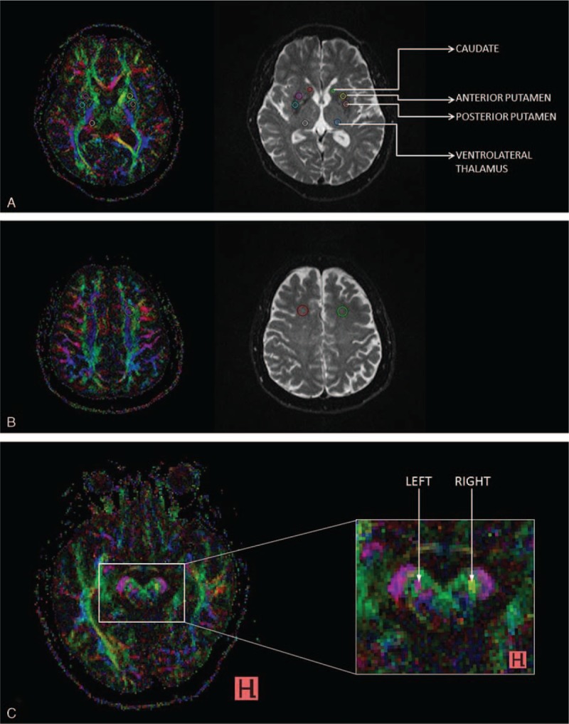
FIGURE 1.

AB colour FA maps (left) and corresponding b0 images (right), depicting placement of regions of interest (ROIs) in the (A) caudate, anterior, and posterior putamen, ventrolateral thalamus, (B) frontal white matter and C, (C) substantia nigra.
