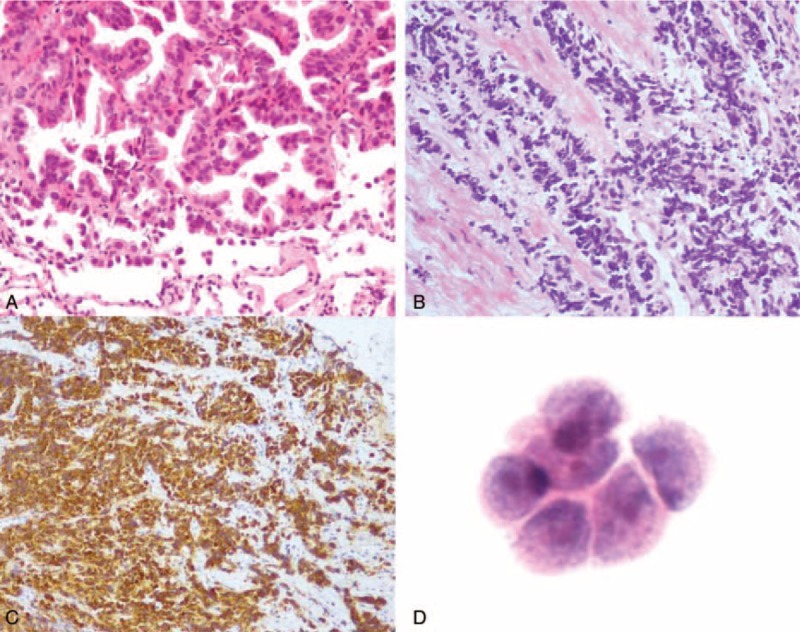
FIGURE 2.

(A) Before treatment, the lung biopsy shows adenocarcinoma, tumor cells are median-sized, with an obvious atypical appearance, abundant cytoplasm, and nuclear division, and are arranged in papillary and acinus along fibrovascular cores. (B) The recurrent tumor contains small cells arrange in a prominent nesting pattern with neuroendocrine morphology and (C) diffuse positive staining for Syn. (D) Adenocarcinoma cells found in cerebrospinal fluid. (A and B) ×300 H&E (hematoxylin and eosin); (C) ×150 Syn IHC (immunological histological chemistry); (D) ×400 H&E.
