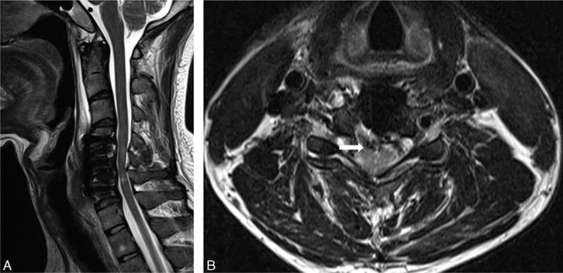
FIGURE 2.

MRI taken 3 weeks postoperatively showed high signal intensity in the spinal cord at C4–C5 and C5–C6 (A), as well as adhesions of the spinal cord to the PLL (arrow) (B). MRI = magnetic resonance imaging, PLL = posterior longitudinal ligament.
