Figure 2.
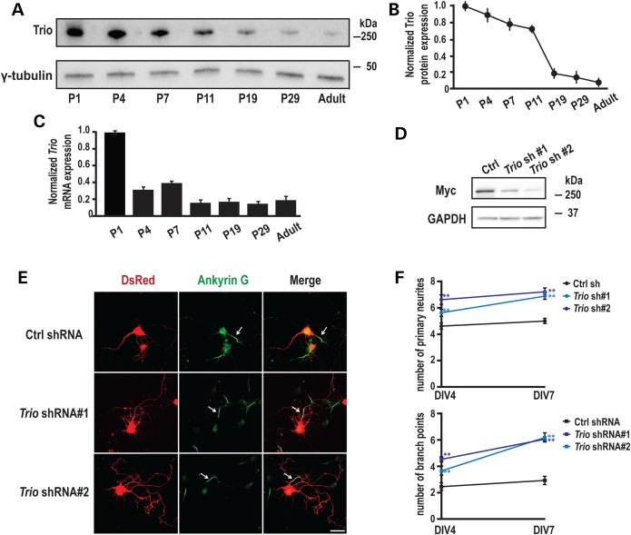
Developmental expression of TRIO and its role in neurite outgrowth. (A) Rat hippocampi were collected at the indicated ages; equal amounts of protein (50 μg) were subjected to western blot analysis and representative blots are shown. (B) Quantification of Trio protein levels at different postnatal ages, normalized to the levels at P1 (n = 3). (C) Quantification of Trio mRNA levels using Q-PCR at different postnatal ages (n = 3). (D) 293 cells co-transfected with vectors encoding MYC-TRIO and Trio shRNA 1 and 2 were harvested after 24 h. Western blot analysis using a MYC antibody demonstrated that both Trio shRNA 1 and 2 efficiently reduced Trio expression. (E) Rat hippocampal neurons nucleofected at the time of plating with a control (scrambled) shRNA or Trio shRNA 1 or 2 were fixed and analyzed at the indicated times. shRNA transfected neurons were identified by DsRed co-expression; axon initial segments were identified by staining for Ankyrin G; representative images at DIV7 are shown. Scale bar, 50 µm. (F) Quantification of primary neurites and branch points at DIV4 and DIV7. Neurons expressing Trio shRNAs exhibited more primary neurites and more branch points than neurons expressing the control shRNA. Five separate experiments were performed; 20–30 neurons per group per experiment were counted. **P < 0.01 with respect to the control (scramble) shRNA vector.

