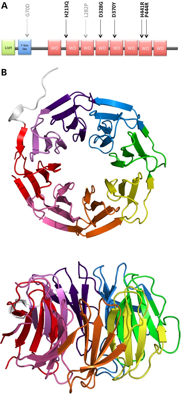
Figure 1.

Structure of TBLR1. (A) Domain structure with location of diagnostic missense mutations. The five new DDD mutations are indicated in black and the two previously published mutations in grey. (B) Three-dimensional β-propeller structure of the WD40 domain from PDB entry 4lg9, top and side views. The eight propeller blades are rainbow coloured, starting with red for the N-terminus through to violet for the C-terminus.
