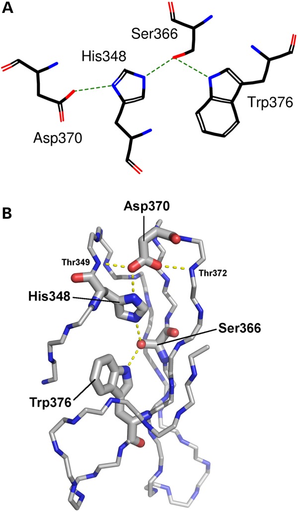
Figure 3.

Representation of the hydrogen-bonding network of the DHSW tetrad. Taken from the fifth WD40 motif in the 3D structure of TBLR1 (PDB entry 4lg9). (A) Schematic representation showing the four sidechains involved: Asp370, His348, Ser366 and Trp376. Hydrogen bonds are shown by the green dotted lines. (B) Three-dimensional representation showing the location and sidechains of the four tetrad residues; the rest of the domain is represented only by backbone atoms N, Cα and C. Potential hydrogen bonds are shown by the dashed lines. Note the importance of the highly conserved Asp370, which can not only hydrogen-bond to the histidine, but also to the backbone of neighbouring strands, helping hold the propeller-blade structure together.
