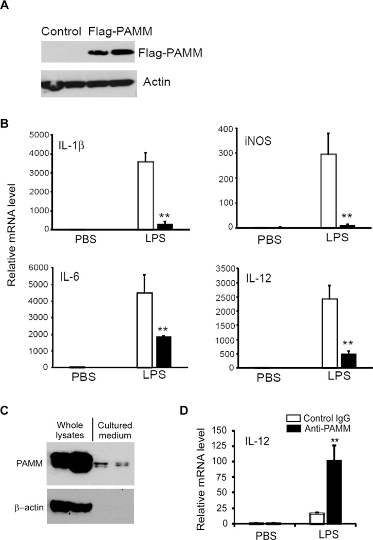
Figure 2. Overexpression of PAMM inhibited inflammatory gene expression in Raw264.7 cells.
(A) Raw264.7 cells were transiently transfected with Flag-PAMM expression plasmid or control plasmid. Cell lysates from transfected cells were analysed by Western blot with anti-Flag antibody. The same membrane was probed by anti-actin antibody and served as a loading control. (B) Transfected Raw264.7 cell were stimulated with or without 1.0 µg/ml LPS for 8 h. Relative mRNA levels of IL-1β, IL-6, IL-12 and iNOS were detected by QPCR. Data represent mean ± S.D., n = 3, **P <0.001 compared with control group. (C) 2-day post-confluent 3T3-L1 cells were incubated with a cocktail of DMI for 10 days to induce differentiation into adipocytes. The cell lysates and cultured media were collected for Western blot analysis of PAMM protein with anti-PAMM. (D) The conditional cultured medium from above experiments was pre-incubated with anti-PAMM (1.0 ng/ml) or control IgG for 1 h, then the medium was added into Raw264.7 cells and incubated for 1 h. Then the cells were stimulated with or without 1.0 µg/ml LPS for 8 h. Relative mRNA level of IL-12 was detected by QPCR. Data represent mean± S.D., n = 3, **P <0.001 compared with control IgG group.

