
Figure 4. Immunochemical analysis of hMSCs stained, after 72 hours, for nuclei, vinculin, and actin cytoskeleton, on the surface of 20wt% RLP hydrogels.
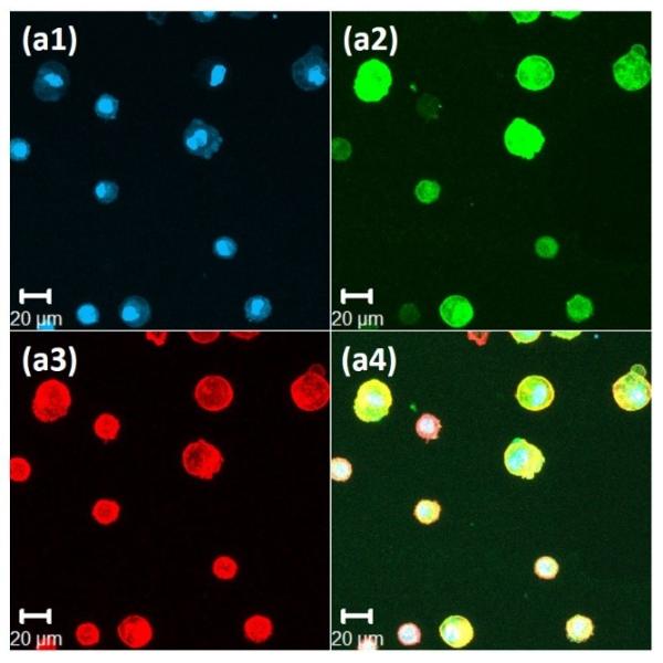
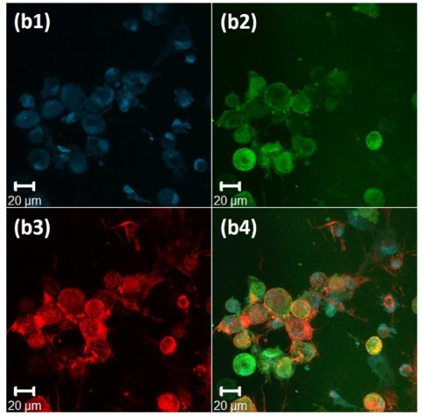
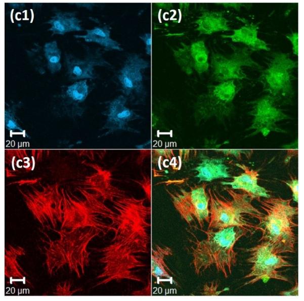
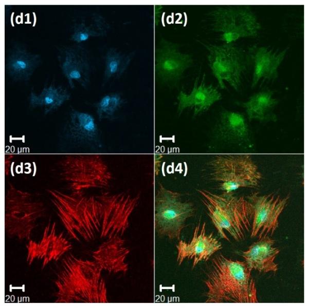
(a) 100% RLP, (b) 100% RLP-RDG, (c) 50% RLP and 50% RLP-RGD and (d) 100% RLP-RGD. All hydrogels were crosslinked at a 1:1 ratio of amine : hydroxyl groups. Data for each hydrogel composition is separated into 4 panels: (1) Cell nuclei counterstained by Draq5 (blue); (2) Focal adhesion sites visualized (green) by treatment with anti-vinculin and a FITC-labeled secondary antibody; (3) F-actin filaments visualized (red) by treatment with TRITC-phalloidin; and (4) the merged image of the triply stained hydrogels (Draq5, vinculin and TRITC-phalloidin).