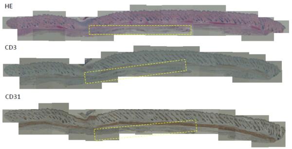
Figure 6. Subcutaneous transplantation of 20wt% RLP hydrogels in Sprague Dawley (SD) rats sacrificed after one week.
Tissue samples were stained and histological images are presented with hematoxylin and eosin (H&E), cluster of differentiation 3 (CD3) and cluster of differentiation 31 (CD31) staining. The yellow box highlights the location of gel transplantation and the remnants of RLP-hydrogel after one week post transplantation.

