
Figure 6. InsG3680 and R1117X Mutant Mice Display Common and Differential Disruptions of Post-synaptic Signaling Complexes.
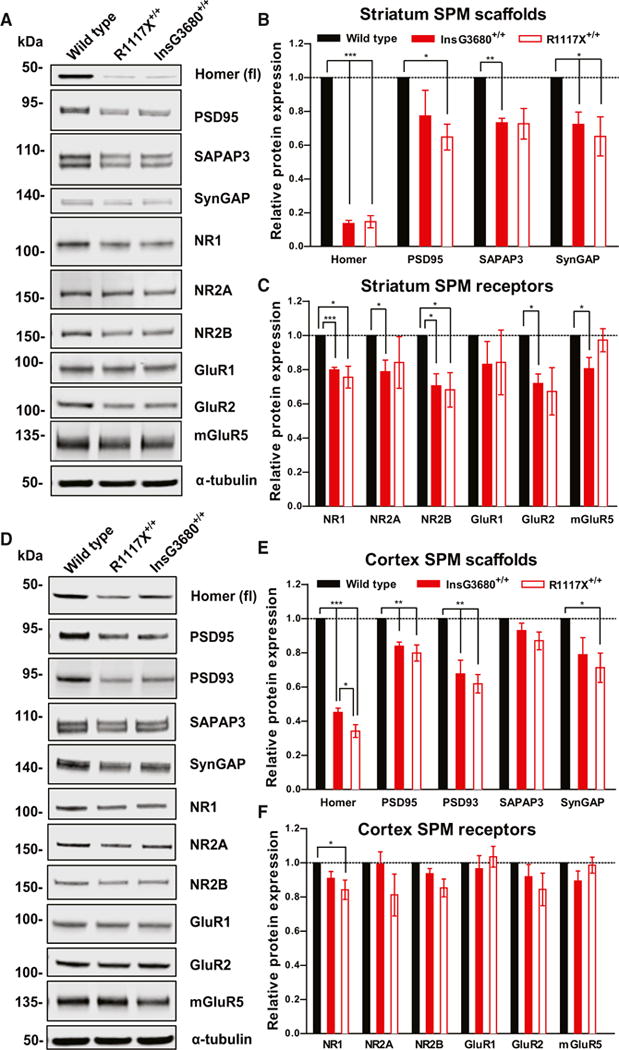
(A) Representative blots for proteins detected by specific antibodies in the striatal SPM fraction from wild-type, InsG3680+/+, and R1117X+/+ mice.
(B and C) Quantification of relative levels of proteins as normalized to tubulin protein expression from striatal SPM. (n = 4–6 samples per protein per genotype, each n being pooled tissue from three mice).
(D) Representative blots for proteins detected by specific antibodies in the cortical SPM fraction from wild-type, InsG3680+/+, and R1117X+/+ mice.
(E and F) Quantification of relative levels of proteins as normalized to tubulin protein expression from cortex SPM. (n = 4–11 samples per protein per genotype, each n being pooled tissue from two mice).
In (B), (C), (E), and (F), data are presented as mean ± SEM. *p < 0.05, **p < 0.01, **p < 0.001; one sample t test.