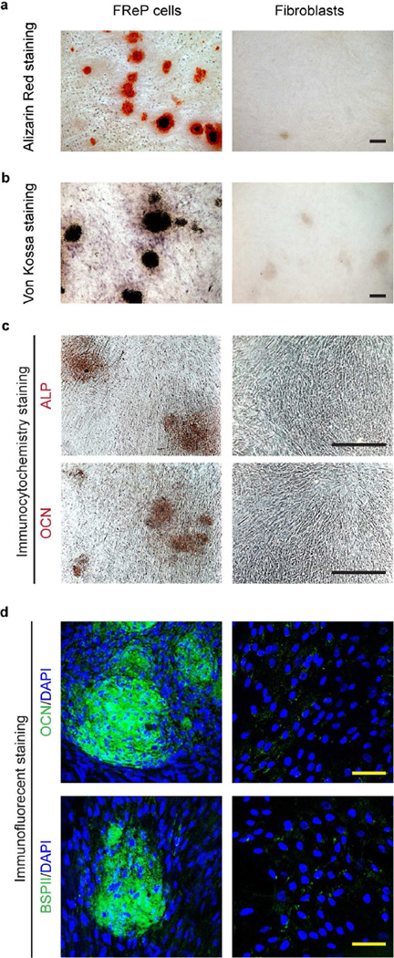
Fig. 2. Osteogenic differentiation of FReP cells after a 4-week cultivation in osteogenic medium in vitro.
(a) Alizarin Red staining, (b) von Kossa staining, (c) immunocytochemistry staining against ALP and OCN, and (d) immunofluorescent staining against OCN and BSPII, respectively. DAPI was used for nuclear staining. Bar = 400 µm (a–c), or 50 µm (d).

