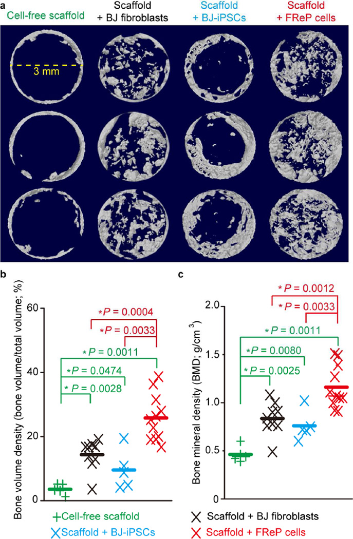
Fig. 5. Radiographic analysis of bone regeneration in critical-sized SCID mouse calvarial defects at week 8 post-implantation.
(a) µCT image of bone regeneration in critical-sized mouse calvarial defects implanted with cell-free scaffold (N = 5), scaffold + undifferentiated BJ-fibroblasts (N = 9), scaffold + BJ-iPSCs (N = 5), and scaffold + FReP cells (N = 11). 5 × 10⁵ cells were seeded on PLGA/HA scaffold and cultured in osteogenic medium 3 days prior to implantation. Images were documented at a resolution of 20.0 µm. (b) Bone volume density and (c) bone mineral density quantification revealed that implantation of FReP cells resulted in significantly more bone formation than other groups in critical-sized SCID mouse calvarial defects at week 8 post-transplantation. *, significant difference revealed by Mann-Whitney test; green stars indicate the significance from the cell-free scaffold; red stars indicate the significance in comparison to scaffold + FReP cells.

