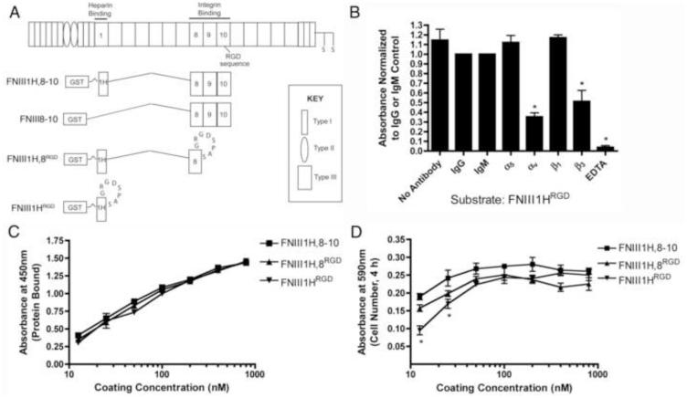
Fig. 1.
Fibronectin matrix mimetics. (A) Schematic representation of a fibronectin subunit and fibronectin fusion proteins. (B) FN-null MEFs were seeded (1.6 × 10⁵ cells/cm2) onto wells pre-coated with FNIII1HRGD (100 nM) in the presence of integrin function-blocking antibodies, isotype-matched control antibodies, or EDTA. Cell adhesion was determined as described in “Methods”. Data are presented as mean fold difference compared to IgG (α5, αv, β3) or IgM (β1) controls ± SEM of 3 experiments performed in triplicate. *Significantly different from +IgG, p < 0.05 (ANOVA). (C) Tissue culture plates were coated with increasing concentrations of proteins. The relative amount of protein bound to wells was determined by ELISA. Data are presented as mean absorbance of triplicate wells ± SEM and represent of 1 of 4 experiments performed. (D) FN-null MEFs were seeded (1.6 × 10⁵ cells/cm2) onto protein-coated wells and allowed to attach for 4 h. The number of adherent cells was determined as described in “Methods”. Data are presented as mean absorbance of triplicate wells ± SEM and represent of 1 of 3 experiments performed. *Significantly different from FNIII1H,8RGD and FNIII1H,8-10, p < 0.05 (ANOVA).

