Fig. 9.
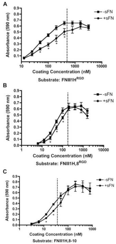
Fibronectin decreases cell-substrate attachment strength during cellular self-assembly. FN-null MEFs were seeded (4 × 10⁴ cells/cm2) onto tissue culture plates pre-coated with increasing concentrations of FNIII1HRGD, FNIII1H,8RGD, or FNIII1H,8-10 and allowed to adhere for 4 h. Cells were then incubated in the absence (−sFN) or presence (+sFN) of soluble fibronectin (50 nM) for an additional 20 h. Centrifugal cell adhesion assays were performed and the number of cells that remained adherent was determined, as described in “Methods”. Data are presented as mean absorbance ± SEM of at least 3 separate experiments, each performed in triplicate. *Significantly different from corresponding ‘−sFN’, p < 0.05 (t-test). The dotted lines denote the initial Cc50 values.

