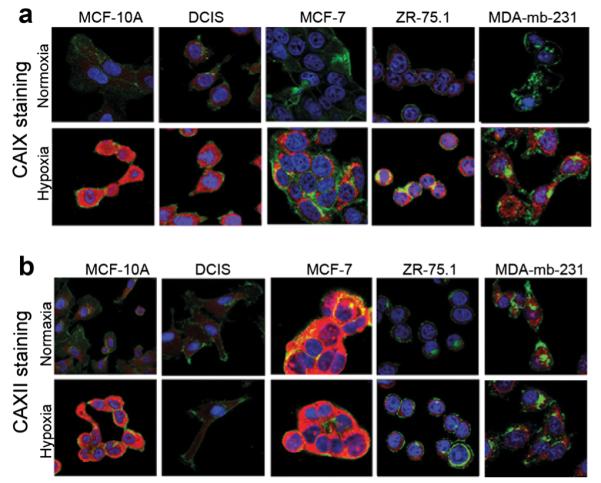
Fig. 2.

a and b ICC staining of breast cancer cells incubated in normoxic and hypoxic conditions using the CA9Ab-680 and CA12Ab-680 probes. Confocal micrographs of cells incubated with the nuclear marker DAPI (blue), the plasma- and cytoplasmic-membrane marker, WGA (green) and CA9Ab-680 or CA12Ab-680 (red). a Stronger CA9Ab-680 staining is observed for all breast cancer cell lines grown in hypoxia relative to normoxia. b Hypoxia-induced CA12Ab-680 staining is observed in MCF-10A and ZR-75.1 cells, and comparable levels of constitutive expression in normoxia and hypoxia are observed in DCIS, MCF-7 and MDA-mb-231 cells. Cytoplasmic staining is observed due to permeabilization of cells after fixation.
