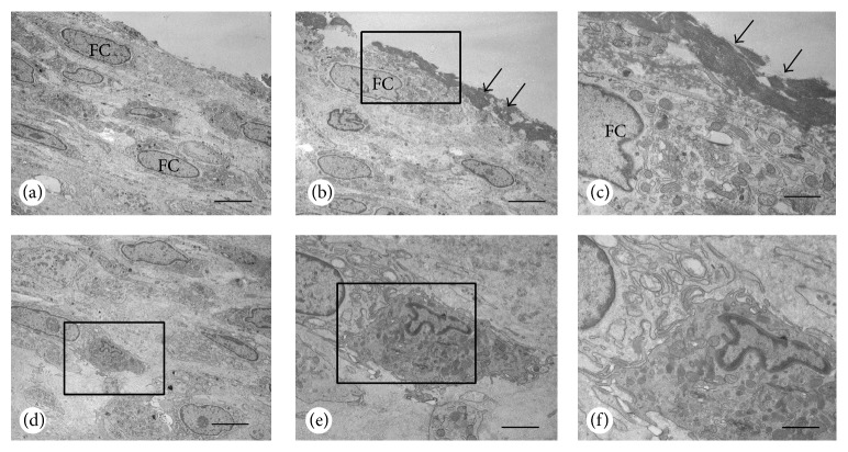
Figure 5.
Electron microscopy of articular cartilage surface with moderate joint destruction. The surface of the articular cartilage was composed of a thick layer of fibroblastic cells (FC, a), which were sometimes covered with electron dense materials (arrows, b). At a higher magnification, electron dense materials overlaid the fibroblastic cells with abundant Golgi apparatus and rough endoplasmic reticulum (c). A macrophage can be seen in the inner portion of the fibroblastic cell layer (d). The macrophage contains many lysosomes and vesicles making contact with fibroblastic cells (e, f). Panel (f) is a magnified image of (e). Bars: (a), (b), (d): 10 μm, (c), (f): 5 μm, and (e): 7 μm.

