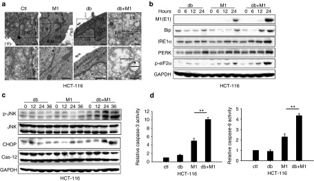
Figure 4.
M1 infection plus db-cAMP treatment induces severe and prolonged ER stress, leading to cell apoptosis. (a) Transmission electron microscopy images (9,700, up; 13,800, down) of HCT-116 cells. The marker indicates the relative size of the ER. N, nucleus. M, mitochondrion. Scale bars, 500 nm. (b) Expression of ER stress markers by western blot. (c) Western blots of p-JNK, Jun N-terminal kinase (JNK), C/EBP-homologous protein, and Caspase-12 in HCT-116 cells treated with db-cAMP, M1, or M1/db-cAMP. (d) Caspase-3 and Caspase-9 activity assays (mean ± SD). HCT-116 cells were plated on 96-well plates and M1 virus was infected for 72 hours in the presence or absence of db-cAMP. **P < 0.01. GAPDH, glyceraldehyde-3-phosphate dehydrogenase.

