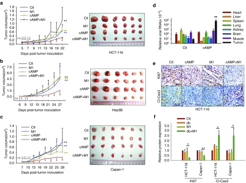
Figure 6.
8-CPT-cAMP plus M1 virus significantly reduces tumor size. (a–c) Measurement of the oncolytic effects of M1 in vivo. Nude mice (NU/NU) bearing subcutaneous HCT-116 (a), Hep-3B (b), and Capan-1 (c) tumors were treated with vehicle, 8-CPT-cAMP (20 mg/kg/day), M1 virus (Hep3B, 5 × 10⁶ plaque forming unit (PFU)/day; HCT-116 and Capan-1, 3 × 10⁷ PFU/day), M1 virus and 8-CPT-cAMP (n ≥ 7). Tumor growth was assessed by tumor volume measurement over time (mean ± SD). At experimental endpoints, mice were anesthetized and sacrificed. Tumors were subsequently dissected and photographed. i.v., intravenously injection (tail vein). **P < 0.01, compared with the combination group. (d–f) In vivo distribution of M1 (d) and intratumoral expression of Ki-67 and cleaved-Caspase-3 (e and f). Immunohistochemistry was performed to analyze the expression of Ki-67 and Cleaved-Caspase-3. Relative protein expressions were quantified with Image-Pro Plus 6.0 (IPP 6.0, MediaCybernetics, Rockville, MD). **P < 0.01, compared with tumor in control group. Scale bars, 50 μmol/l.

