Figure 2.
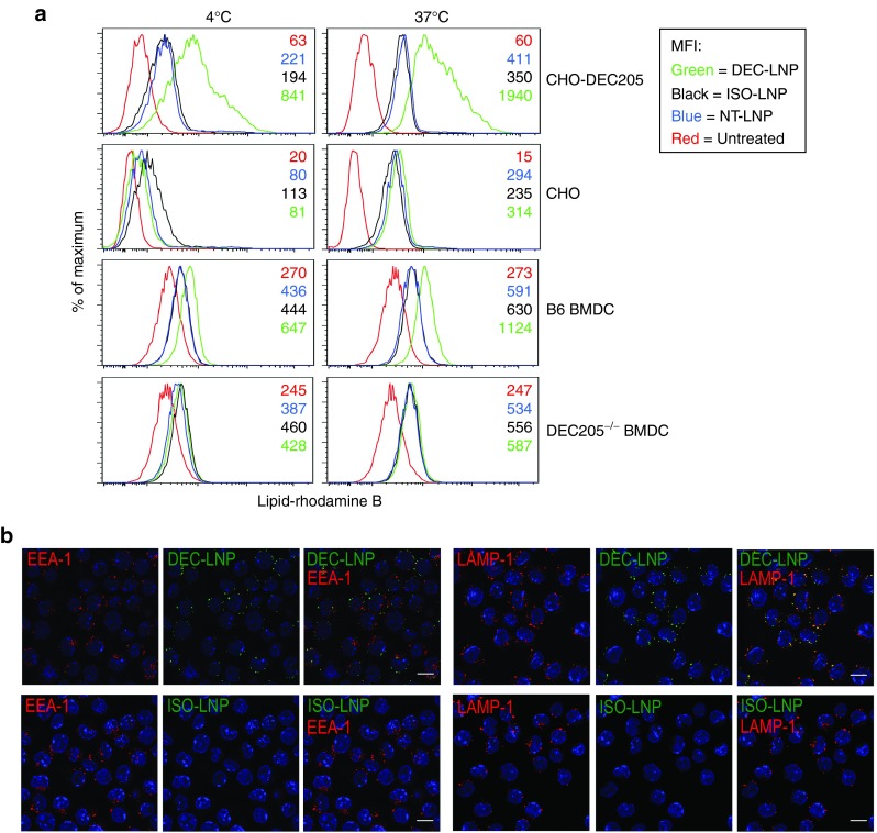
DEC-lipid nanoparticles (LNPs) are taken up by DEC205+ cells and colocalize to lysosomes. (a) CHO-DEC205 (top panel) and parental CHO (second panel), B6 bone marrow-derived dendritic cell (BMDC) (third panel) and DEC205–/– BMDC (fourth panel) were incubated with DEC-LNPs (green), ISO-LNP (black), NT-LNP (blue) or were left untreated (red). Cells were incubated at 4 °C (left panel) or 37 °C (right panel) for 30 minutes, washed and analyzed by flow cytometry. Numbers in panels denote mean fluorescence intensity. (b) DEC-LNPs and ISO-LNPs containing fluorescently labeled siRNAs were incubated for 1 hour, 37 °C with A20 cells (DEC205+). Following fixation and permeabilization, cells were stained with EEA-1 and LAMP-1 and cellular localization of LNPs determined by confocal microscopy. DEC-LNP, ISO-LNP = green; EEA-1, LAMP-1 = red; DAPI = blue. Images acquired with 63× objective. 10 μm scale bars are shown in the merged images.

