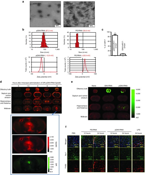
Figure 2.
pGNVs have a better capacity for carrying RNA without toxicity. RNA loaded pGNVs (pGNV/RNA) and PEI-RNA were purified by ultracentrifugation. (a) Sucrose-banded pGNV/RNA and PEI-RNA were visualized and imaged by electron microscopy. (b) Size distribution (top panel) and Zeta potential (bottom panel) of pGNV/RNA) or PEI/RNA were analyzed using a ZetaSizer. (c) Loading efficiency of miR17-Dy547 was determined using a fluorescence microplate reader (EX/Em = 530/590 nm) and expressed as % = (miR17-DY547 in pGNV/RNA or GNVs)/total RNA used for loading × 100%. Images (a, b) and data (c, n = 5) are representative of at least three independent experiments. (d) Intranasal administration of pGNV/RNA-Syto60 results in localization to the brain. Syto60-labeled RNA (20 µg, red) carried by DIR-labeled pGNVs (green) was administered intranasally to C57BL/6j mice. At different time points post intranasal administration, the brain was cut sagittally, and the ventral sides of cut brain were placed against the scanner for imaging using the Odyssey laser-scanning imager. Enlarged images are shown at the bottom. (e) DIR-labeled GNVs or pGNV/RNA was administered intranasally to C57BL/6j mice. At 12 hours post intranasal administration, the brain was cut sagittally, and the ventral sides of cut brain were placed against the scanner for imaging using the Odyssey laser-scanning imager. (d,e) Representative sagittal images from the center of the brain (n = 5). (f) Intranasal administration of pGNVs does not induce brain macrophages. pGNV/RNA or PEI/RNA were administered intranasally to C57BL/6j mice. Mice were sacrificed 12 or 24 hours after intranasal administration of pGNV/RNA or PEI/RNA. C57BL/6j mice were i.p. injected with bacterial lipopolysaccharide (2.5 mg/kg) or PBS as a control and sacrificed at 12 and 24 hours postinjection as a control. Brain tissue sections were fixed as described in the Materials and Methods section. Frozen sections (10 µm) of the anterior part of the brain were stained with the antimicroglial cell marker Iba-1 (green color) or macrophages (red). Slides were examined and photographed using microscope with an attached camera (Olympus America, Center Valley, PA). Each photograph is representative of three different independent experiments (n = 5). Original magnification: ×40.

