Figure 3.
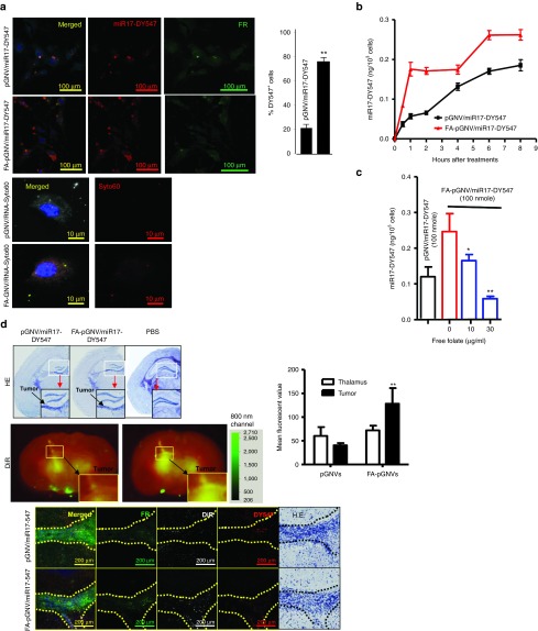
Folate receptor-mediated uptake of FA-pGNVs. GL-26-luc cells were cultured in the presence of Dylight547-labeled miR17 or Syto60-labeled RNA carried by FA-pGNVs (FA-pGNV/miR17-Dy547, FA-pGNV/RNA-Syto60) or by pGNVs (pGNV/miR17-DY547, pGNV/RNA-Syto60). (a) Representative images of cells (n = 3) were taken at 2 hours after addition of FA-pGNVs/miR17-Dy547, pGNVs/miR17-DY547, FA-pGNV/RNA-Syto60, or pGNV/RNA-Syto60 using a confocal microscope at a magnification of ×200 (top panel) or ×600 (bottom panel) and quantified by counting the number of DY547+ cells in five individual fields in each well. % of DY547+ cells was calculated based on the number of DY547+ cells/numbers of FR+ cells × 100. The results are presented as the mean ± SEM. **P < 0.01. (b) At different time points, post incubation at 37 °C, transfection efficiency of FA-pGNV/miR17-Dy547 or pGNV/miR17-DY547 was analyzed by measuring fluorescent density using a microplate reader at Ex/Em = 530 /590 nm. (c) Before adding to GL-26-luc cultures, FA-pGNV/miR17-Dy547 (100 nmol) was premixed with different concentrations of FA, and then, GL-26-luc cells were cultured in the presence of FA premixed with FA-pGNV/miR17-Dy547 for 2 hours. The effects of folate on the transfection efficiency of FA-pGNV/miR17-Dy547 were analyzed by measuring fluorescent density using a microplate reader at Ex/Em = 530/590 nm. Data in a–c are the mean ± SEM of two experiments (n = 5). (d) FA-pGNV/miR17-Dy547 more efficiently targeted brain tumor. 2 × 10⁴ GL26-luc cells per mouse were injected intracranially in 6-week-old wild-type B6 mice. Five-day tumor-bearing mice were then treated intranasally with FA-pGNV/miR17-Dy547 in PBS or pGNV/miR17-Dy547. FA-pGNV/miR17-Dy547 in PBS or pGNV/miR17-Dy547 (red representing miR17 labeled with Dylight 547, 20 µg of miR17 carried by pGNVs) was administered intranasally into C57BL/6j mice. Results of hematoxylin and eosin (HE) staining showing tumor tissue as indicated by arrows (top panel). DIR dye-labeled FA-pGNV/miR17-Dy547 or pGNV/miR17-Dy547 (second panel from the top, the results represent the mean ± SEM of three independent experiments, bar graph). HE-stained brain sections of GL-26-luc tumor-bearing mice (the first column from right) or miR17-Dy547 (red) or anti-folate receptor (FR) antibody stained (green) brain tumor sections and adjacent area of mice treated with the agents listed (third and fourth panel from the top). Original magnification: ×20. Data represent at least three experiments with five mice/group.

