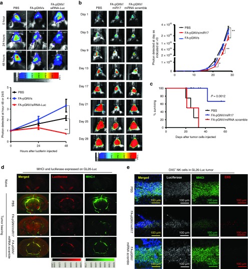
Figure 4.
FA-pGNV/miR17-Dy547 treatment prevents the growth of in vivo injected brain tumor cells. 2 × 10⁴ GL26-luc cells per mouse were injected intracranially in 6-week-old wild-type B6 mice. Fifteen-day tumor-bearing mice were then treated intranasally on a daily basis with FA-pGNVs/siRNA-luc or FA-pGNVs/siRNA scramble control. The mice were imaged on the hours as indicated in a. (a) A representative photograph of brain tumor signals of a mouse from each group (n = 5) is shown (top panel). In vitro time course of bioluminescent signal generated by GL26-Luc cells is shown (bottom panel). The results are based on two independent experiments with data pooled for mice in each experiment (n = 5) and are presented as the mean ± SEM. **P < 0.01. (b) Mice were intracranially injected with GL26-luc and treated every 3 days for 21 days beginning on day 5 after tumor cells were implanted. The mice were imaged on the days as indicated in the labeling of b. (b) A representative photograph of brain tumor signals of a mouse from each group (n = 5) is shown (left). In vitro time course of bioluminescent signal generated by GL26-Luc cells is shown (right panel). The results are based on two independent experiments with data pooled for mice in each experiment (n = 5) and are presented as the mean ± SEM; *P < 0.05, **P < 0.01. (c) Percent of FA-pGNVs/miR17, FA-pGNVs/scramble miRNA, or PBS mice surviving was calculated. One representative experiment of four independent experiments is shown (n = 5 females per group). (d) Results of anti-luciferase and MHCI staining or (e) antiluciferase/MHCI/DX5 staining of brain tumor sections and adjacent area of mice treated with the agents listed. Original magnification: ×20. Data represent at least three experiments with five mice per group.

