Figure 2.
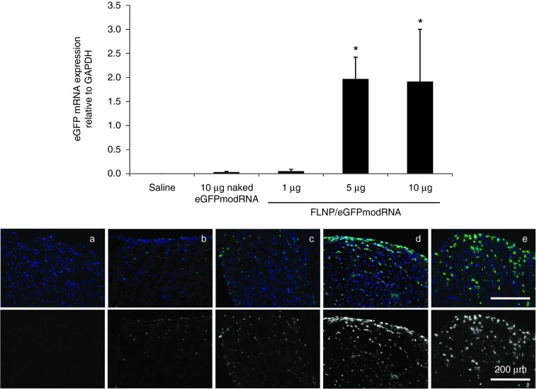
Dose response of intramyocardial injection of formulated lipidoid nanoparticles (FLNP)/eGFPmodRNA in rats. Top panel: Expression of eGFP mRNA measured by real-time polymerase chain reaction in rat myocardium 20 hours after intramyocardial injection of: FLNP with 1, 5, or 10 μg of eGFPmodRNA, and either saline or 10 μg of naked eGFPmodRNA as controls. Values represent mean (±SD) eGFP mRNA expression levels relative to GAPDH, n = 3 rats per group. *P ≤ 0.02 versus naked eGFPmodRNA. Bottom panel: Immunofluorescence micrographs of corresponding frozen tissue sections of rat myocardium for (a) saline-only, (b) 10 μg naked eGFPmodRNA, and (c–e) FLNP/eGFPmodRNA at 1-μg (c), 5-μg (d), or 10-μg (e) doses. Sections incubated with anti-GFP antibody (green), and nuclei stained with DAPI (blue). Upper row shows pseudo-colored images with merged green and blue channels. Lower row shows only the green channel in grayscale. Scale bar = 200 μm for all panels. DAPI, 4',6-diamidino-2-phenylindole.

