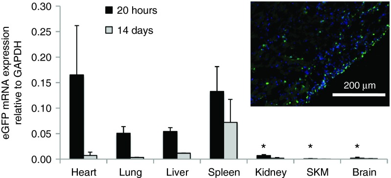
Figure 5.
Intracoronary delivery of FLNP/eGFPmodRNA via left ventricle injection with temporary aortic cross-clamping in the rat. Bar graph displays the biodistribution of eGFP mRNA in organs harvested 20 hours (black bars, n = 4) and 14 days (gray bars, n = 2) after intracoronary delivery of formulated lipidoid nanoparticles (FLNP) with 10 μg of eGFPmodRNA. Values represent mean (±SD) expression levels of eGFP mRNA measured by real-time PCR relative to GAPDH. SKM, skeletal muscle. *P < 0.005 relative to heart at 20 hours; no statistical analysis was done for the 14-week samples. Inset: GFP expression in rat myocardium collected at 20 hours postdelivery, demonstrated by immunofluorescence of frozen tissue section incubated with anti-GFP antibody (green), and DAPI (blue). Scale bar = 200 μm. DAPI, 4',6-diamidino-2-phenylindole.

