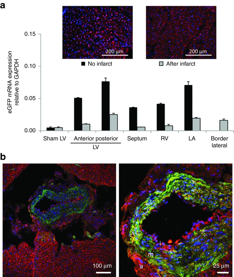
Figure 7.
Intracoronary delivery of FLNP/eGFPmodRNA in a pilot large animal study in healthy and diseased pigs. (a) Expression of eGFP mRNA in healthy and diseased (48 hours post-MI) pig myocardium 20 hours after intracoronary injection of FLNP with 500 μg dose of eGFPmodRNA, comparing expression levels in different regions of the heart. LA, left atrium; LV, left ventricle; RV, right ventricle. Insets: Immunofluorescence of posterior LV frozen tissue sections from healthy (left inset) and postinfarct (right inset) hearts show GFP expression (red), and nuclei stained with DAPI (blue). Scale bars = 200 μm. (b) Immunofluorescent microscopy studies show GFP expression (red) colocalizes with α-smooth muscle actin-positive smooth muscle cells in the media (m) and also with cells in the intima (i) and adventitia (a) layers of the coronary vessel wall, which is surrounded by GFP-positive myocardial tissue. Scale bars = 100 and 25 μm, as shown. DAPI, 4',6-diamidino-2-phenylindole.

