Figure 2.

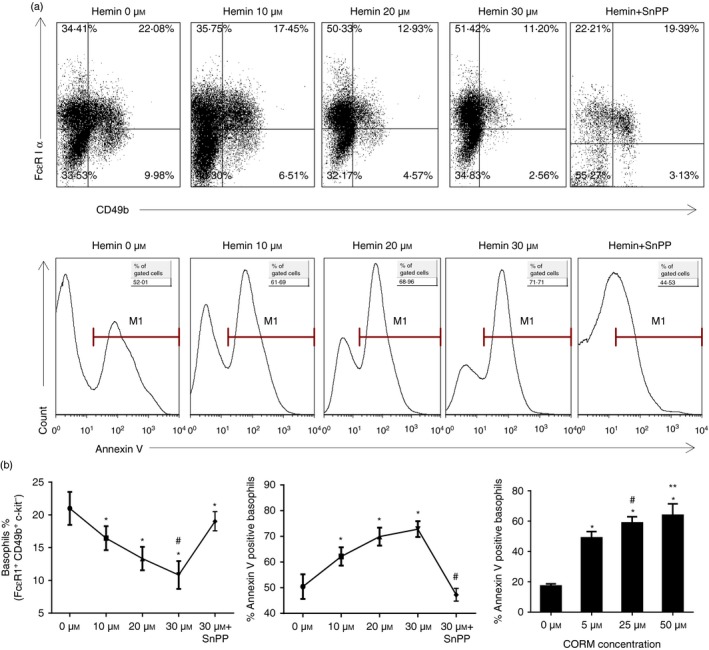
HO‐1 and exogenous CO inhibited basophil maturation and promoted their apoptosis in vitro. Bone marrow cells from normal BALB/c mice were cultured for 10 days with IL‐3. Different doses of hemin (0, 10, 20 and 30 μmol/l) or CORM (0, 5, 25, 50 μmol/l) were added. In the hemin plus SnPP group, 30 μmol/l hemin and 30 μmol/l SnPP were used respectively. Flow cytometry analysis was performed to detect the percentage and apoptosis of basophils. (a) Flow cytometeric analysis of the percentage of basophils in culture under different concentrations of hemin and hemin plus SnPP (gated on c‐kit− basophil‐enriched population; upper panel); Annexin V expression in basophils under different concentrations of hemin and hemin plus SnPP (gate on Fcε RI α + CD49b+ PI − subset; low panel). (b) Left, the percentage of basophils (gated on Fcε RI α + CD49b+ c‐kit− subset (*P < 0·05, versus 0 μmol/l group; #P < 0·05, versus hemin+SnPP group, respectively); Middle, the percentage of Annexin V expression in basophils (gate on Fcε RI α + CD49b+ PI − subset; *P < 0·05, versus 0 μmol/l group; #P < 0·05, versus 10, 20 and 30 μmol/l group, respectively); Right, the percentage of Annexin V expression in basophils (gate on Fcε RI α + CD49b+ PI − subset; (*P < 0·05, versus 0 μmol/l group, respectively; #P < 0·05, 25 μmol/l group versus 5 μmol/l group; **P < 0·05, 25 μmol/l group versus 50 μmol/l group). Data are representative of three independent experiments.
