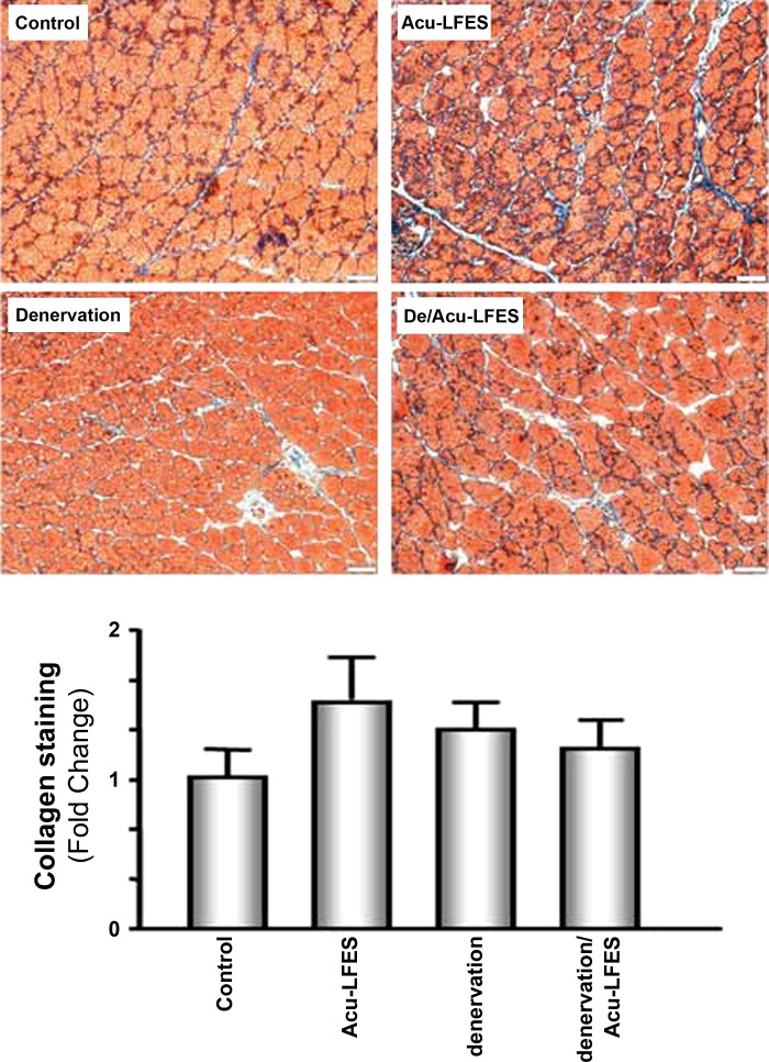
Fig. 5.
Collagen appears in the muscle of Acu-LFES mice. Muscle samples were collected 14 days after acupuncture. Muscle frozen sections were stained with Masson modified IMEB trichrome to detect collagen deposition in the gastrocnemius muscle of normal control, Acu-LFES, denervation, or denervation/Acu-LFES mice. The red color indicates muscle fiber; the blue indicates collagen; and the black indicates nuclei. The bar graph compares the area (μm2) of blue staining per 500 muscle fibers in each group expressed as a fold change from levels in control mice, which is represented by 1 (bars: means ± SE; n = 6/group; no statistically significant change).

